Best Retina Surgery in Ahmedabad

What is retinal vein occlusion in Ahmedabad?

Occlusion (blockage) of a retinal vein is a common cause of sudden painless reduction in vision in older people. Sanjivani Eye Hospital is one of the leading Retina Surgery specialist in Ahmedabad. Offering low cost Retina Surgery in Ahmedabad.

The retina is the thin membrane that lines the inner surface of the back of your eye. Its function is similar to that of the film in a camera. Blockage of one of the veins draining blood out of the eye causes blood and other fluids to leak into the retina, causing bruising and swelling as well as lack of oxygen. This interferes with the light receptor cells (cells responsible for vision) and reduces vision.

The condition is uncommon under the age of 60 but becomes more frequent in later life.
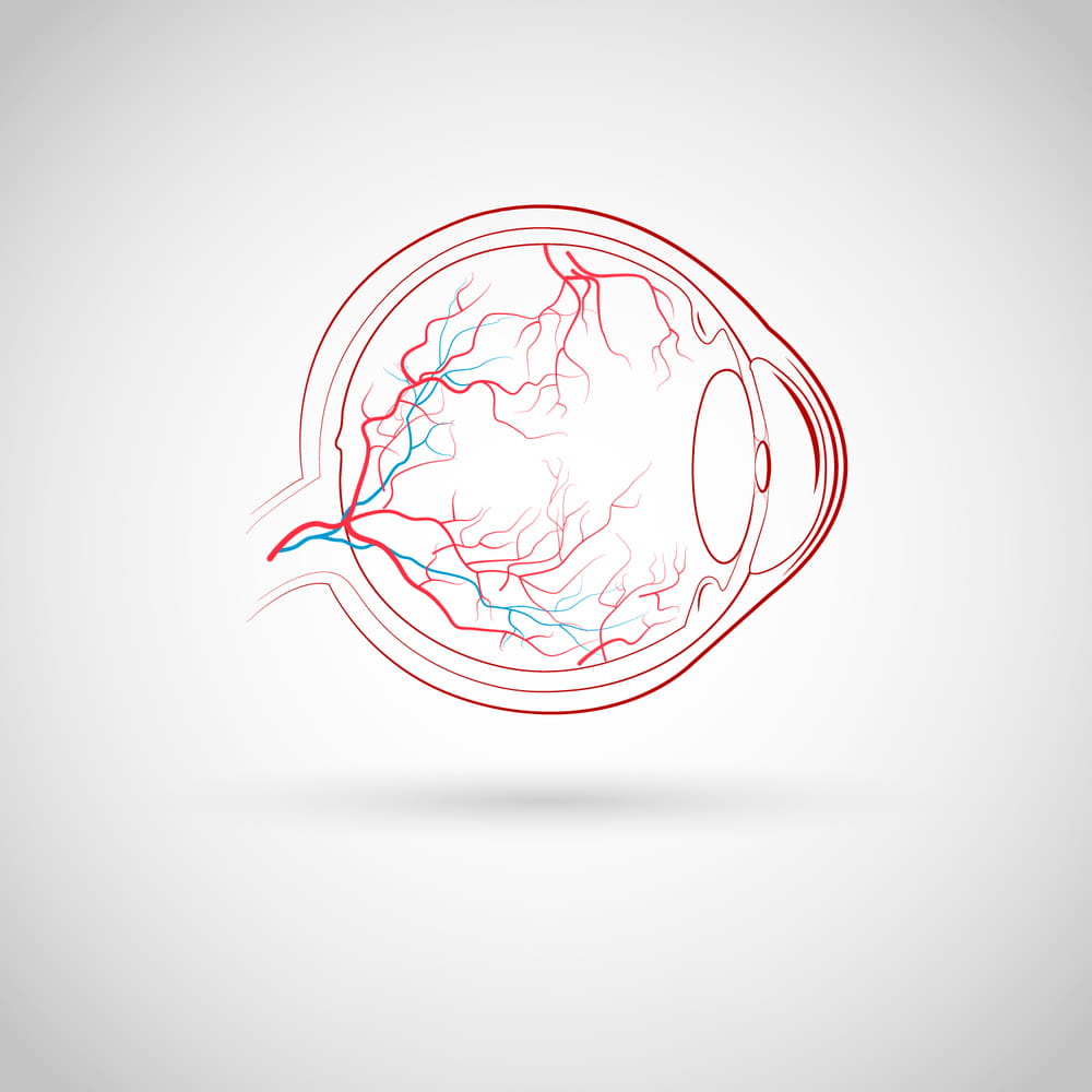
Retina Surgery in Ahmedabad

THERE ARE TWO TYPES OF RETINAL VEIN OCCLUSION IN AHMEDABAD

Branch retinal vein occlusions are due to blockage of one of the four retinal veins, each of which drains about a quarter of the retina

Central retinal vein occlusion is due to blockage of the main retinal vein, which drains blood from the whole retina

It causes painless loss of vision, In general, visual loss is more severe if the central retinal vein is blocked.

What causes retinal vein occlusion in Ahmedabad?

Retina Surgery in Ahmedabad - Top 10 best retina surgery specialist in Ahmedabad, Gujrat, India.

A blockage forms in the vein, usually due to a blood clot, and obstructs the blood flow. The exact cause is unknown, but several risk factorsmake the condition more likely. These include

  • High blood pressure
  • High cholesterol
  • Glaucoma
  • Diabetes
  • Smoking
  • Certain rare blood disorders

Diagnose Retina Surgery in Ahmedabad

Retina specialists in Ahmedabad are ophthalmologists that have completed training (including a one- or two-year fellowship) after their three year ophthalmology residency to specialize in diseases and conditions related to the vitreous and retina specialist surgery in Ahmedabad. Ophthalmologists in Ahmedabad are physicians who specialize in medical and surgical eye care(Retina Surgery Specialist in Ahmedabad).

How do you diagnose retinal vein occlusions?

Ophthalmoscopy The changes caused by RVO may be seen by examination of the retina with an instrument called an ophthalmoscope. We are one of the top-rated retina surgery in Ahmedabad, Gujarat.

Optical coherence tomography (OCT) This is a high definition image of the retina taken by a scanning ophthalmoscope (computer with special light and lens). These images can determine the presence of swelling and edema by measuring the thickness of your retina. The doctor will use OCT images to objectively document the progress of the disease throughout the course of your treatment. In this Situation Meet with retina specialist doctor in ahmedabad.

Fluorescein angiography This is a test procedure in which a dye that is injected into a vein in the arm travels to the retinal blood vessels. Photographs on a special camera allow the physician to see the vessels and detect leakage, neovascularisation or lack of blood supply

Why do people lose vision from retinal vein occlusions?

Macular edema. The macula is the small, central area of the retina that allows sharp, detailed vision, such as that necessary for reading. Blood and fluid leaking into the macula due to block in the vein imparing blood drainage cause swelling, a condition called macular edema, which causes blurring and/or loss of vision.

Neovascularization. RVO can cause the retina to develop new, abnormal blood vessels, a condition called neovascularization. These new vessels may leak blood or fluid into the vitreous, the jelly-like substance that fills the inside of the eye. Small spots or clouds, called floaters, may appear in the field of vision. With severe neovascularization, the retina may detach from the back of the eye.

Neovascular glaucoma. New blood vessels in certain parts of the eye can cause pain and a dangerous increase in pressure inside the eye which is quite resistant to conventional treatment.

The complications of RVO, especially if they are not treated, can lead to irreversible loss of vision .

Retina Surgery Hospital in Ahmedabad

What is the treatment for RVO?

Since there is no cure for either BRVO or CRVO, the focus of management is on the following: The evaluation and treatment of underlying risk factors (e.g., diabetes or hypertension), leading to improved treatment of systemic problems and prevention of heart attack and stroke. The evaluation and treatment of complications such as macular edema and neovascularization, leading to preservation of vision.

MACULAR EDEMA

Laser photocoagulation: Laser has been shown to be effective in improving macular edema in BRVO but not CRVO. A usually painless laser beam is directed through the pupil toward the affected region of the macula. Complications are uncommon with the new subthreshold and micropulse laser treatment.

Anti-VEGF injections: In both BRVO and CRVO, abnormally high levels of vascular endothelial growth factor (VEGF) occur, promoting both macular edema and neovascularization. Anti-VEGF medications that counteract VEGF are injected directly into the vitreous through the sclera (white of the eye). The eye is numbed before each injection and the doctor closely monitors each patient's condition following the injection.

Two medications currently being used, Lucentis® / Accentrix (ranibizumab) and Avastin® (bevacizumab), have both been successful in reducing the edema and improving vision in both BRVO and CRVO. A third medication, Eylea® (aflibercept), is effective for treating edema secondary to RVO.

A common treatment approach is to start with 2-3 monthly injections and continue as needed over the next several months.

Complications are rare, but can include a) infection (endophthalmitis) b) retinal detachment and c) vitreous hemorrhage, all of which are related to the injection itself and not specifically to the medications.

Although only Lucentis and Eylea are approved by the FDA for treatment, Avastin is also commonly used in this manner, and all are thought to be effective and equally safe for the eye.

Steroids: Steroids are another type of medication that can decrease macular edema. Similar to Lucentis, Eylea and Avastin, the steroid is injected directly into the vitreous through the sclera. Two of the most commonly used steroids are preservative-free Triamcinolone Acetonide Suspension and Ozurdex (a dexamethasone implant). The primary difference between them is that the effect of Ozurdex is longer in the eye than triamcinolone (6 months versus 2 months). The complication of steroid induced glaucoma and cataract progression is much more with thriamcinolone than Ozurdex.

Repeat injection are required frequently over the course of the disease.

NEOVASCULARIZATION

Laser photocoagulation: Laser is the best way to control and decrease neovascularization in either BRVO or CRVO. A stronger laser is applied to the affected region of the retina. Mild peripheral vision loss can sometimes occur.

Vitreous hemorrhage and tractional retinal detachment: If the retina specialist is unable to perform the laser treatment due to either a vitreous hemorrhage or retinal detachment, a vitrectomy will be recommended. A vitrectomy is an outpatient surgical procedure performed in an operating room that involves removal of the vitreous from the inside back of the eye through three openings made in the sclera using a microscope and several small instruments. Now a days Micro incision sutureless vitrectomy (MIVS) is done using 25 and 27 Gauge instruments .

NEOVASCULAR GLAUCOMA

Treatment includes control of Intraocular pressure (IOP) via topical, oral and systemic medications. Also it is required to treat the underlying retinal disease which is causing tremendous amounts of VEGF via PRP laser, AntiVEGF injections and Cryopexy. In some cases antiglaucoma surgery in form of Trabeculectomy with MMC or Valve surgery is required.

As with any treatment, it is critical that patients have a thorough discussion with their retina specialist regarding all of the treatment options, including the risks, benefits and alternatives, before deciding the appropriate course of action.

Top 10 Retina Specialist in Ahmedabad, Best Retina Doctor in Ahmedabad, Best Retina Specialist in Gujarat, Best Retina Specialist in India, Retina Eye Specialist in Ahmedabad, vitreo retinal surgeon in ahmedabad